
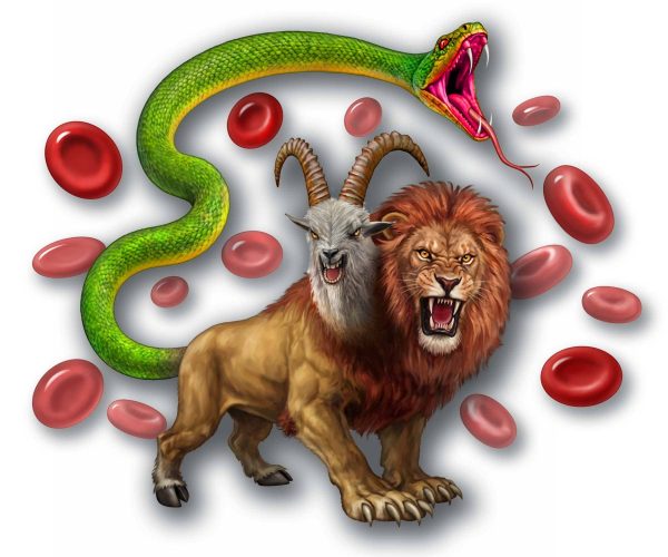
If you are a fan of Greek mythology, the word “chimera” may be familiar to you. In this context, it refers to a hybridized lion that exhales fire and has a second goat head emerging from its upper thoracic vertebrae. Oh, and let’s not forget that a large and menacing serpent has replaced the lion’s tail. Two horns protrude from its mane, and it barks like a dog. When used in a literary context, it can refer to anything sinister, unimaginable, unthinkable, or even mesmerizing (personally, we lean toward ‘greatly disturbing’!).
For a pharmaceutical chemist, however, the word has a different meaning. In this context, a chimeric molecule is an engineered drug made by linking distinct compounds into a new chemical entity, combining or enhancing the functions of its components to achieve a specific therapeutic goal. An example is linking a monoclonal antibody to a cytotoxic drug that targets and destroys cancer cells.
Ask a biologist about the meaning of chimera, and they’d likely describe a lifeform, whether flora or fauna, having cell lines of completely different forms. In this case, it’s as if somehow different species merged to form a hybridized creature. An example is when a male deep-sea anglerfish embeds itself in the female to form a single organism. Maybe the Greeks were thinking like contemporary biologists!
Military wounded who received blood transfusions
But chimeras need not be dramatic hybrids of different species, engineered molecules, or divergent cell lines. Sometimes the foreign presence is far more subtle — a small population of cells from another individual, quietly persisting within a host who remains entirely unaware. This is microchimerism: a low-level genetic coexistence, hidden in plain sight within the bloodstream. And as it happens, one of the most direct pathways for foreign cells to enter the body is through something medicine has relied on for decades — a blood transfusion.
It took 60 years after our modern blood banking industry was founded, but in 1977, investigators first reported that, following a blood transfusion, donor white cells proliferated in some recipients, then disappeared over about 10 days. Fast forward to the beginning of the 21st Century, and investigators discovered that up to 15% of those who received blood after traumatic injury had persistent survival of the donor’s white cells, a phenomenon they termed “transfusion-associated microchimerism,” designated TA-MC. As more observations of the phenomenon were reported, it became clear that lineages of donor white cells often persisted in the transfusion recipient, and in some, proliferated over the post-transfusion years. This strongly suggested that new cell lines had engrafted (perhaps “seeded” would be apropos?) into the recipient. Different cell lines, different DNA, occupying the same person.
While researching papers for another topic, we came across an interesting one that piqued our interest regarding blood administration. Published in the high-impact journal Transfusion, researchers assessed if TA-MC persisted beyond a couple of years, pursuing the question in an interesting way.
Subjects were recruited following a call for volunteers printed in an issue of Disabled American Veterans’ magazine, with a circulation of over 1.75 million. It asked readers who had received a blood transfusion in World War II, the Korean War, or the Vietnam War because of injuries they had received to consider participating in a study. They excluded those who had undergone a more recent organ or stem cell transplantation or a blood transfusion to avoid confounding influences.
Blood and medical histories were obtained in 163 (all male) combat veterans, all over the age of 60 at the time of the study, who met the inclusion criteria, and a control group, matched for war service, age, and absence of ever receiving a transfusion. We won’t go into the sophisticated technologies used (e.g., polymerase chain reaction assay, nucleic acid testing) or details of the statistical analysis. What was found was that transfusion in this group of combat-injured men commonly resulted in microchimerism (MC) persisting for 60 years or more, demonstrating that the donor’s DNA was, in fact, proliferating in the recipient for decades.
Leukoreduction to the rescue? Maybe not!
This got us to thinking about the many times we’ve administered leukoreduced blood for one reason or another. Recall that this process removes as many white blood cells (leukocytes) as possible, decreasing transfusion reactions such as TRALI and lessening the risk of transmitting cytomegalovirus. If white cells are the culprits, then surely leukoreduction would mitigate against MC. We went looking in the literature and came across a study, also in Transfusion, that unfortunately demonstrated persistent MC in civilian trauma victims transfused with leukoreduced blood.
What about transfusion in children?
Once again, we drilled down and found a 2024 study in children who had been transfused with leukoreduced, gamma-irradiated blood. The authors lament that pediatric transfusions are rare, that a significant volume of blood is required to test for MC, and that research in children faces logistical hurdles. These greatly complicate its study. While they detected MC in several patients, they urged more studies to define the risks. But a question remains: are very young children particularly vulnerable to the consequences of a foreign DNA populating their bodies?
What is the risk of persistent microchimerism? Much ado about nothing?
Recall that during pregnancy, maternal and fetal blood mix. A bidirectional movement of cells from the mother, the fetus, and the placenta results in what’s termed trans-migratory fetal cells (TMFC) that can be detected for decades throughout the body of the mother following normal birth. This is what is known as “natural” MC, as it results from the course of a normal pregnancy. MC in this context might prove to be either deleterious or beneficial.
Deleterious effects might include a predisposition to future preeclampsia, possibly some cancers, and autoimmune disorders (recall, 80% of patients with autoimmune disorders are female). Potential benefits might include an empowered immune system, protection against some cancers, and enhanced repair of injured tissues. We lean towards the latter, as it may help explain the longer life expectancy of females!
But what may be more onerous is the seeding of foreign DNA from donor to recipient, which is known to disseminate throughout the body’s tissues and organs. Iatrogenic seeding might occur with transfusion, stem cell infusion, and organ transplantation. From the literature we found, largely focusing on maternal/fetal MC, the long-term effects are uncertain and difficult to study. Hypotheses range from being beneficial to benign to harmful. The most relevant harms involve the immune system, particularly autoimmune disorders and increased cancer risk.
Decision making
Should we consider the potential impact of microchimerism when deciding whether to transfuse blood? Should the risks of MC that might be associated with transfusion be part of the blood consent with the patient? We think not to both, any more than we should be concerned that if there are space aliens out there, they might occupy Earth and hold us hostage (or worse!).
Our current knowledge is nascent at best and awaits further refinement for research and clinical observation to provide decision-making recommendations; a process likely to take years to unfold. For those who have received an iatrogenic seeding that persists as MC, at this point, we are cautiously optimistic that their “hybridization” is largely benign, with little suggesting proof of any seriously deleterious effects. Stay tuned, we will continue to track the evidence stream as it emerges!
Related Articles